By Abigail E. Ward and Miranda Bell-Tilcock

This is no ordinary witch’s brew. It’s one part of the recipe to study thiamine deficiency in our California Central Valley Chinook salmon (Oncorhynchus tshawytscha) populations. In 2019, hatcheries noticed an eerie and shivering change in juvenile Chinook salmon. Offspring were laying on their side at the bottom of tanks, swimming in corkscrew motion (see video below), or not surviving at all. In other words, for many juvenile salmon, they were not quite dead, only slightly alive.

After much deliberation and study by pathologists from USFWS, CDFW and UC Davis, it was diagnosed that salmon were experiencing thiamine deficiency (lacking in Vitamin B1). When fish received a bath in thiamine, individuals went from mostly dead to fully alive!
But now, we are left with a major question: how did these fish became thiamine deficient in the first place? And if this is being seen in our hatchery fish, what about the fish in the wild? To understand thiamine deficiency in salmon, we need to understand the past, present, and future using an array of different samples.

Eyes – To understand the past, we need look no further than deep into salmon eyes (right). The lens within the eye is an onion like structure that can be peeled to reveal multiple layers. Each of these layers represents a different period of the salmon’s life and takes approximately 1-2 months to form, telling us more about what the fish was consuming in the ocean, in essence generating a retrospective diet journal (Bell-Tilcock 2021). For example, one extant hypothesis surrounding thiamine deficiency is that salmon were eating too many anchovies in the ocean. Anchovies are rich in thiaminase, which can break down thiamine in a fish if consumed in large enough quantities. We are also looking to use the lens to better understand diet of Chinook salmon more generally during years when thiamine deficiency occurred.

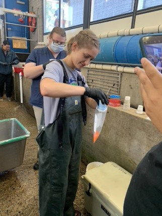
Muscle – To bridge the past to the present, we extract a muscle plug from the salmon (left). When salmon migrate back into rivers to spawn, they are no longer feeding. They use all remaining energy reserves for spawning migration and to produce progeny. Similar to salmon eye lenses, stable isotopes in muscle tissue can reveal dietary patterns. Unlike eye lenses, however, this allows us to understand recent diet, typically just the last few months, before they returned from the ocean. However, muscle tissue also provides clues into present conditions when analyzed for thiamine. Combined, muscle tissues allow us to see how salmon are storing thiamine in tissues, and compare concentrations to other tissues in their bodies.

Blood – To understand the present, we extract vials of blood from freshly spawned salmon (right). Just like for humans, we can analyze the blood of salmon to obtain a profile of vitamins and micronutrients circulating their system. Blood Thiamine concentrations will further contextualize patterns seen in tissues, eyes and other organs.
Liver – In addition to the blood, we also extract a slice of liver to understand the present. The liver is a key factor in metabolism, and thiamine concentrations provide another indication of thiamine deficiency. In comparing liver concentrations to those of the muscle and eggs, we can test for severity of deficiency since the liver conserves thiamine more than other tissue types.
Eggs – And finally to understand the future, we examine eggs (progeny) from the recently departed (left). With a small sample of eggs, we can understand how much thiamine is being passed from the mother to her progeny. Egg thiamine concentrations also help mitigate against thiamine deficiency in future populations by understanding how patterns change across different runs, at different rates, and different times. Hatchery managers can then use this information to adjust treatments of both thiamine injections for adults and thiamine baths at fertilization. It also helps researchers cue in on which fish we may want to target for eye lens and muscle research to better understand life-history variations.
Taken together, these samples collectively help us better understand the threat thiamine deficiency poses to our Chinook salmon populations, and aides in developing new research methods for future studies surrounding this complicated problem. Collaborations with multiple labs and agencies is critical in solving the wicked problems that fish face in today’s climate and human-dominated landscapes.
Researching these parts and pieces may make us feel like witches and wizards at times, mixing potions and casting spells on these fish to magically heal them. But in the end, our hope is that each ingredient listed here would lead us to answers to ensure our Central Valley Chinook are not living mostly dead, but rather, mostly alive.
Further reading:
https://www.latimes.com/environment/story/2021-01-26/king-salmon-vitamin-deficiency-sacramento-river
https://www.sciencefriday.com/segments/salmon-eyes/
https://californiawaterblog.com/2021/10/24/fish-eyes-the-hidden-diet-journal/
https://californiawaterblog.com/2020/10/31/the-freezer-of-horrors/
Bell-Tilcock, M.N., C.A. Jeffres, A.L. Rypel, M. Willmes, R.A. Armstrong, P. Holden, P.B. Moyle, N.A. Fangue, J.V.E. Katz, T.R. Sommer, J.L. Conrad, and R.C. Johnson. 2021. Biogeochemical processes create distinct isotopic fingerprints to track floodplain rearing of juvenile salmon. PLoS ONE 16(10): e0257444 .
Bell-Tilcock, M., C.A. Jeffres, A.L. Rypel, T.R. Sommer, J.V.E. Katz, G. Whitman, and R.C. Johnson. 2021. Advancing diet reconstruction in fish eye lenses. Methods in Ecology and Evolution 12: 449-457.
Leave a Reply